The preclinical laboratory staff of Iris Pharma is trained for microdissection of animal ocular samples. The most common are: palpebral or bulbar conjunctiva, nictitating membrane, extra ocular muscles, lacrimal gland, Harderian gland, nasal lacrimal duct, cornea, aqueous humor, iris, ciliary body, lens, vitreous, retina, choroid, sclera, optic nerve and eyelids.
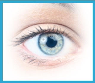
For more details on the location of these ocular samples and also the essential components of the human eye's optical system, please move mouse over the Diagram (.jpg) ).
).
.jpg "eye structures")
Screenshot of human eye anatomy on McGraw-Hill website - Image courtesy of The McGraw-Hill Companies, Inc.